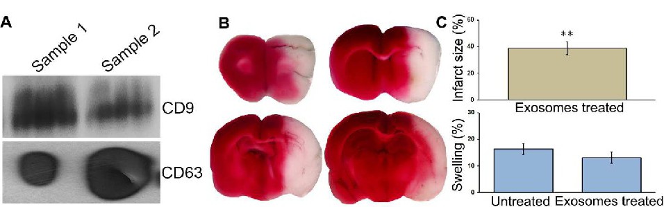
Fig. 2. Effect of treatment with exosomes on infarct size and swelling. (A) Detection of exosomal markers (CD9 and CD63) expression by immunoblot analysis. (B) Representative TTC stained images of rat coronal brain sections at one-day reperfusion subsequent to a two-hour focal cerebral ischemia in rats. (C) Bar graphs represent the percent infarct size and ipsilateral hemisphere swelling in exosomes and/or untreated rats. Histograms and error bars indicate the mean and the SEM, respectively. n = 6. Quantitative data of infarct size and swelling obtained from exosomes treated rats at one-day reperfusion was compared with our previously published data of untreated, ischemia-induced rats [33]. **p<0.01 vs. untreated ischemia-induced rats euthanized at one-day reperfusion.